Dividing cancer cell, fluorescent microscopy - Vídeo de acervo HD
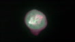
Timelapse fluorescence microscopy video of a dividing cancer cell in 3D tissue culture. The total length of the sequence is 3 hours. The cell is expressing a fluorescently-tagged version of a protein (EB1) that binds to growing microtubule ends and also localizes to the spindle poles. This highlights the highly dynamic microtubule cytoskeleton in the spindle, and also shows astral microtubules extending toward the cell periphery. The different colors represent different focal planes giving a more 3D impression. In the beginning of the video it can be seen how the duplicated centrosomes move around the cell nucleus and then form the poles on opposite ends of the mitotic spindle apparatus. Although chromosomes themselves are not labeled, they can be seen as darker shadows in the cytoplasm. The cell eventually divides apparently without all chromosomes having aligned correctly in the middle of the spindle, leading to aneuploidy, one of the hallmarks of cancer. A slowed down version of this clip can be seen as K007/4486


COMPRAR UMA LICENÇA
Informe quando, onde e como deseja usar este arquivo para receber ofertas de preço personalizadas.
DETALHES
Crédito:
ID Criativo:
1432329434
Tipo de licença:
Direitos prontos
Coleção:
Photolibrary Video
Tamanho máximo do arquivo:
1920 x 1080 px - 271 MB
Duração do clipe:
00:00:21:16
Data do upload:
Info sobre autorização:
Não requer autorização
Masterizado em:
QuickTime 8-bit Photo-JPEG HD 1920x1080 25p
Categorias:
- Célula Cancerígena,
- Câncer - Tumor,
- Telefone celular,
- Biologia,
- Oncologia,
- Esquisito,
- Repetição - Conceito,
- Ampliação,
- Time Lapse,
- Cromossoma,
- Microtúbulo,
- Divisão celular,
- Fundo preto,
- Metástase,
- Pesquisa,
- Anáfase,
- Bilro,
- Citoesqueleto,
- Condição,
- Doença,
- Experimento,
- Filme - Imagem em Movimento,
- Formato de Alta Definição,
- Horizontal,
- Imagem a cores,
- Metafase,
- Micrografia,
- Mitose,
- Mitótico,
- Prófase,
- Tecnologia sem Fio,
- Velocidade Alta,